
Healthy correspondent, BBC East Midlands
 Bbc
BbcDigital imaging of suspicious skin cancers has considerably reduced waiting times for diagnosis and treatment in Nottinghamshire.
Clinical photographers in certain parts of the county now see patients referred by general practitioners generally in a few days, rather than having to wait what was sometimes weeks to see a consultant for an initial meeting.
Imaging includes the use of artificial intelligence smartphone software (IA), which then sees photos sent to a consultant dermatologist to assess, without the need to meet the patient.
Sherwood Forest Hospitals NHS Foundation Trust said that the approach was to release specialists to focus on surgery.

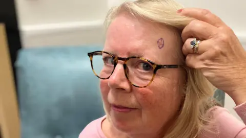
Fiona Hayward-Lyon, from Farndon in Nottinghamshire, is one of the nearly 2,000 patients seen by the trust with suspected skin cancer which has benefited from faster access to the diagnosis.
The service was fully introduced in June 2024 and a year later, was described as a “great success” by the trust.
Ms. Hayward-Lyon had become concerned about a lesion on her forehead.
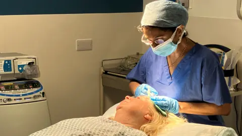
The 63-year-old meeting for photos was organized within three days of his GP view, and it took a little more than four weeks to obtain the operation, which took place in October.
“I had a red imperfection on my forehead for a while and I suddenly noticed that he was rising,” she said.
A dermatologist then examined his images remotely.
Only three percent of patients under the care of the trust require a face-to-face follow-up meeting after initial photography, and some like Ms. Hayward-Lyon require surgery.
In his case, a basal cell carcinoma – a type of skin cancer – has been diagnosed requiring elimination.
She said, “I didn’t expect to be seen so quickly. I can now move on and be a little more careful in the sun.”
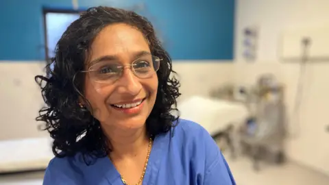
The consultant dermatologist, Dr. Ritu Singla, who treated Ms. Hayward-Lyon, said that the photography service had allowed doctors to reassure patients earlier if they did not have cancer.
“We can exclude a lot of benign [non-cancerous] Lesions, which are most of the cases, “she said.
“It also allows us to start treatment earlier for patients where cancer has been diagnosed.”
Dr. Singla said: “Patients are more aware of skin cancer these days, [but] At the same time, in the aftermath of the pandemic, we had long waiting lists.
“We prioritized, but some patients were waiting for months for treatment.”
There is a national NHS objective for 96% of skin cancer cases to be treated with 31 days of decision to deal with.
Before the introduction of the photography service, the trust reached 72% in the first quarter of 2023-24.
The latest figures show that 100% of patients in February 2025 were treated within the target time.
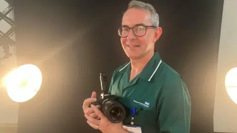
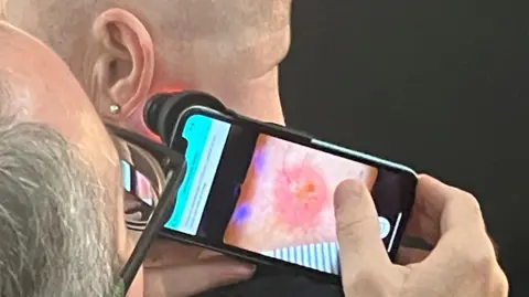
Clinical photographer Jason Randall says that he uses a special polarized light, a device called Dermatoscope, to help produce high resolution images.
He said: “This allows the camera to see in the first layer of the skin not clearly visible to the naked eye and especially the edges of the lesion.”
The greater use of technology is one of the themes of the new NHS government plan at the NHS, which seeks to improve efficiency, productivity and results for patients.


